VariPrep is your solution for MALDI Matrix Application:


  • 5x faster than your common glass sublimator: sample preparation in as little as 2 minutes!
  • robust and comparable results by easy workflow adaptation
  • simplicity for all levels of experience enables operator flexibility
  • free trial for riskless evaluation


Get your quote here


Stay tuned for new products and updates by subscribing to our newsletter below!

EXPERIENCES


Lots and lots of DHB

So we used VariPrep to apply MALDI matrix DHB via sublimation to a cooled tissue sample. We subsequently did data acquisition without in between recrystallization. MSI-results were achievable, yet left room for improvement. It was not yet clear where and how. We wanted to see what the deposited matrix looks like after the laser scanning at 5µm pixel size, thus went for SEM imaging. As the sample made its way there by post, it appeared to have encountered...percussive impact.


Result: The glass slide was  damaged, and the layer of deposited matrix was shattered, debris fell of the sample tissue, consequently partially uncovering what was underneath. Nothing we would have done purposefully at this point, but I appreciate the accidental insights. All in all, we might have been a little too generous with the DHB, as the sublimation yielded with 80% efficiency! A lot more than previously experienced with glass sublimators. Here is the SEM image of the matrix on tissue.


What can be seen in the image quadrants is:
 

  • from top left the tissue with partially grown in crystals

  • at the top right a consistent layer of crystals grown atop of the tissue (not ripped off)

  • the bottom left shows a layer of matrix, that has been scanned for MALDI-MSI at size of 5µm for pixels and raster

  • at the bottom right is a portion of the deposited matrix without laser ablation marks, (reminds me of  cauliflower)
     

The bottom right and top right differ in their appearance, i.e. the cauliflowerish top layer is smoother than the just-above-the-tissue layer in this image. My interpretation is that the matrix, if too thick, forms layers that are at least laterally coherent enought to fall off as such if impacted. Especially once the matrix crystals were melted by the laser and baked together. Also, for the laser ablation marks, while they did in fact give molecular data, their depth does not effectively reach the tissue. If the top surface is closer to the tissue, the amount of sample that will be ionized might increase, ultimately resulting in higher signals for the analyte and not intra-matrix-ionization.


What are your ideas on this? Let's have a chat about it!


All the best

Christian

VISION

Life science research needs to improve participation and effectiveness!


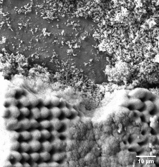
Methods in analytical laboratories often require a variet of tools and chemicals. For a variety of niche applications, there has been a demonstration of a way a particular step can be done. Yet the pursuit often stopped before optimization. This also applies to MALDI-MSI sample preparation. MALDI-MSI is a key technology on the verge of personalized medicine. In our opinion, there are perceived limits which have to be overcome for unfolding this technologys potential.


We aim to make research more affordable in terms of investment, training and workforce to boost its democratization.

WHO WE ARE

Christian - Founder

Electronics and crafting have been my creative tools since youth. Living, multifaceted technology suits me, which is why I decided to study Biology and Sustainable Process Engineering and Biotechnology. I then started my professional life as a certified software tester in medical imaging and subsequently worked as a research assistant in the field of chemical analysis. I applied my motivation for solution development to custom challenges in the life science environment.